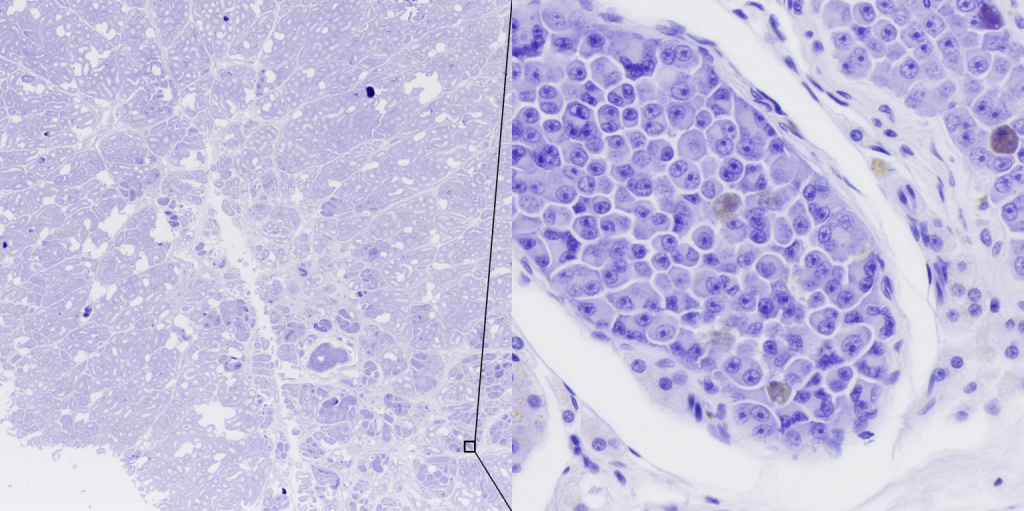
The sixth generation teams have been working on their startups since the beginning of the new year through participation in the Startup 101 bootcamp within the Erasmus+ project Cogsteps and through SPOCK workshops, and now it is time to introduce them. The first team we are presenting is Digicyte, whose goal is to digitalize tissue samples with the help of the Marvin scanner to facilitate early diagnosis from biopsies—something particularly important in tumor detection.
Diagnosis from biopsies, that is, from samples of tissue, cells, and bodily fluids, is still largely based on manual, slow, and often subjective examination under a microscope. Early and accurate diagnosis is crucial, especially for tumors. However, microscope slides must be examined at very high magnifications, where it is often impossible to thoroughly review the entire sample. In addition, doctors are human, and humans have good days and bad days and are prone to fatigue and distractions. All of this leads to variability in the analysis of tissue and cell samples.
The development of deep learning algorithms, or “artificial intelligence,” has offered a glimmer of hope that things could finally change. However, most institutions and clinics still cannot afford scanners that would allow them to digitalize tissue samples. While digitalization has become ubiquitous in our everyday lives, in medicine it remains “reserved” only for the largest laboratories and institutions in the West. Digitalization is key because it “opens the door” to using computational algorithms, and therefore artificial intelligence, in image analysis. Without digital images, there is no artificial intelligence.
Aside from digitalization, another major barrier to the widespread use of AI in tissue sample analysis is the large number of images needed to train neural networks. Annotating medical images is not only time-consuming – it also requires a deep understanding of the data, i.e., pathology. High expertise combined with time demands in medicine leads to high annotation costs, which are often unsustainable. Without a tool that can automatically and accurately annotate large quantities of images, we will never achieve widespread use of artificial intelligence in medicine.
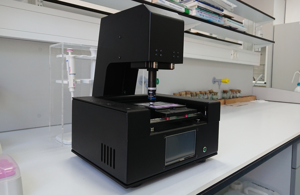
Digitalization and artificial intelligence will revolutionize medical diagnostics. But to make this happen, the focus must be not only on developing AI applications, but also on developing an affordable scanner that enables widespread use of these solutions. For this reason, we developed Marvin – a tissue sample scanner that is more than an order of magnitude (>10×) more affordable than existing solutions on the market. This was achieved through the development of numerous state-of-the-art algorithms capable of compensating for simpler hardware and, in general, providing much better functionality (e.g., fully automatic, “one-button” scanning).
Marvin is essentially a motorized high-resolution microscope (250 nm). It was developed entirely from scratch, inspired by 3D printers. By using small yet very fast neural networks, Marvin has been given a “human-like” understanding of tissue – that is, the ability to automatically locate and scan tissue samples, eliminating the need for a technician and thus reducing operational costs. This makes it possible for small clinics or even regions lacking adequate specialists, such as certain parts of Croatia – to offer their patients expert analysis that would otherwise be unavailable or significantly harder to obtain.
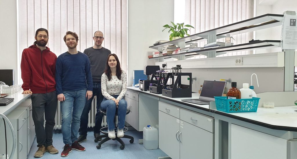
Our team currently consists of four people with mutually complementary expertise. Dora Machaček and Krunoslav Vinicki come from the field of veterinary medicine and understand very well the needs and challenges doctors face in everyday practice. Dora primarily works on image data processing and AI application development aimed at quantifying different cells of interest in scanned tissues. Krunoslav is responsible for developing the scanner’s software. His main goal is to use smart algorithms and fast neural networks to reduce hardware costs, thereby enabling widespread digitalization in pathology.
Dominik Sremić is a robotics engineer who brings broad industrial knowledge to the project. While studying at the Faculty of Mechanical Engineering and Naval Architecture, he received two Rector’s Awards and independently designed and built several versions of 3D printers and electric scooters. Within the team, he is responsible for hardware development—the electronics and mechanics of the tissue scanner.
Leo Obadić is the newest member of our team. He graduated from the Faculty of Electrical Engineering and Computing and has several years of experience in developing complex software solutions.
We have been working together on this project for two years, during which we have built four scanner versions and numerous state-of-the-art algorithms. As a development-focused team, during the first two years we concentrated exclusively on technical solutions and unfortunately neglected all other aspects of launching and running a startup. Our encounter with Matija Srbić and the SPOCK incubator was actually our first real contact with the startup world. Although the sixth generation of the SPOCK incubator has only just started, we can already say that it has drastically accelerated the development of our startup. Through lectures and expert consultations offered by the SPOCK program, we have acquired essential knowledge and sped up development, and thanks to information about competitions we could apply for, this year we won the Startup Factory competition and received our first significant development funding.
We are aware that many challenges still lie ahead, but we have no doubt that with SPOCK, we will overcome them much more easily and quickly. By the end of the program, we see ourselves as a well-organized team with fully developed and market-ready hardware and software solutions for the digitalization of medical samples, having developed our first AI application and with our first collaborations in human medicine already established.